


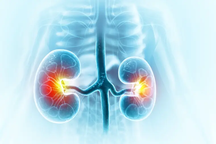
In ureteroscopy, a stone in the ureter (the tube that carries urine from the kidney to the bladder) is reached with a thin camera passed through the urinary tract, then broken up with a laser and removed. No skin incision is made.
A ureteral stone usually causes pain in the side, colicky pain spreading to the groin, blood in the urine, nausea and frequent urination. Small stones may pass on their own. Stones that do not pass, cause infection or make the kidney swell need treatment.
How Is the Procedure Done?
Under general or regional anaesthesia, a thin ureteroscope is passed through the urethra up to the stone. The stone is broken into small pieces with a holmium laser and the fragments are removed with special graspers. If necessary, a temporary tube (DJ stent) is placed in the ureter.
Advantages
- No incision is made
- The stone is treated under direct vision
- The hospital stay is usually short
Afterwards
You will be given advice on fluid intake and diet to reduce the risk of new stones; if needed, stone analysis and a metabolic assessment are carried out. If a DJ stent was placed, it is removed with a simple procedure at the end of the agreed period.
Frequently asked questions
How long does ureteroscopic stone treatment take?
When can I go back to work after ureteroscopy?
What should I keep in mind while I have a DJ stent?
How long does it take for a ureteral stone to pass?
When is the DJ stent removed?
Can ureteral stones come back?
Last updated:
